© 2014 WebMD, LLC. All rights reserved.
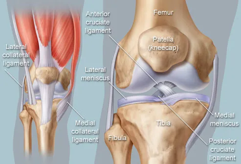
The knee is one of the largest and most complex joints in the body. The knee joins the thigh bone (femur) to the shin bone (tibia). The smaller bone that runs alongside the tibia (fibula) and the kneecap (patella) are the other bones that make the knee joint.
Tendons connect the knee bones to the leg muscles that move the knee joint. Ligaments join the knee bones and provide stability to the knee:
- The anterior cruciate ligament prevents the femur from sliding backward on the tibia (or the tibia sliding forward on the femur).
- The posterior cruciate ligament prevents the femur from sliding forward on the tibia (or the tibia from sliding backward on the femur).
- The medial and lateral collateral ligaments prevent the femur from sliding side to side.
Two C-shaped pieces of cartilage called the medial and lateral menisci act as shock absorbers between the femur and tibia.
Numerous bursae, or fluid-filled sacs, help the knee move smoothly.
Knee Conditions
- Chondromalacia patella (also called patellofemoral syndrome): Irritation of the cartilage on the underside of the kneecap (patella), causing knee pain. This is a common cause of knee pain in young people.
- Knee osteoarthritis: Osteoarthritis is the most common form of arthritis, and often affects the knees. Caused by aging and wear and tear of cartilage, osteoarthritis symptoms may include knee pain, stiffness, and swelling.
- Knee effusion: Fluid buildup inside the knee, usually from inflammation. Any form of arthritis or injury may cause a knee effusion.
- Meniscal tear: Damage to a meniscus, the cartilage that cushions the knee, often occurs with twisting the knee. Large tears may cause the knee to lock.
- ACL (anterior cruciate ligament) strain or tear: The ACL is responsible for a large part of the knee’s stability. An ACL tear often leads to the knee “giving out,” and may require surgical repair.
- PCL (posterior cruciate ligament) strain or tear: PCL tears can cause pain, swelling, and knee instability. These injuries are less common than ACL tears, and physical therapy (rather than surgery) is usually the best option.
- MCL (medial collateral ligament) strain or tear: This injury may cause pain and possible instability to the inner side of the knee.
- Patellar subluxation: The kneecap slides abnormally or dislocates along the thigh bone during activity. Knee pain around the kneecap results.
- Patellar tendonitis: Inflammation of the tendon connecting the kneecap (patella) to the shin bone. This occurs mostly in athletes from repeated jumping.
- Knee bursitis: Pain, swelling, and warmth in any of the bursae of the knee. Bursitis often occurs from overuse or injury.
- Baker’s cyst: Collection of fluid in the back of the knee. Baker’s cysts usually develop from a persistent effusion as in conditions such as arthritis.
- Rheumatoid arthritis: An autoimmune condition that can cause arthritis in any joint, including the knees. If untreated, rheumatoid arthritis can cause permanent joint damage.
- Gout: A form of arthritis caused by buildup of uric acid crystals in a joint. The knees may be affected, causing episodes of severe pain and swelling.
- Pseudogout: A form of arthritis similar to gout, caused by calcium pyrophosphate crystals depositing in the knee or other joints.
- Septic arthritis: An infection caused by bacteria, a virus, or fungus inside the knee can cause inflammation, pain, swelling, and difficulty moving the knee. Although uncommon, septic arthritis is a serious condition that usually gets worse quickly without treatment.
Knee Tests
- Physical examination: By examining the location of knee pain and looking for swelling or abnormal movement, a doctor gathers information about potential causes of damage or stress on the knee.
- Drawer test: With the knee bent, a doctor can pull (anterior drawer test) and push (posterior drawer test) the lower leg while holding the foot stable to check the stability of the ACL and PCL knee ligaments.
- Valgus stress test: Pushing the calf outward while holding the thigh stable, a doctor can check for injury to the medial collateral ligament (MCL). Pushing the calf inward (varus stress test), a doctor can look for injury to the lateral collateral ligament (LCL).
- Knee X-ray: A plain X-ray film of the knee is typically the best initial imaging test for most knee conditions.
- Magnetic resonance imaging (MRI scan): Using high-energy magnetic waves, an MRI scanner creates highly detailed images of the knee and leg. An MRI scan is the most-often used method of detecting ligament and meniscal injuries.
- Arthrocentesis of the knee (joint aspiration): A needle is inserted into the joint space inside the knee, and fluid is drawn out. Various forms of arthritis may be diagnosed through knee arthrocentesis.
- Arthroscopy: A surgical procedure that allows examination of the knee with an endoscope.
Knee Treatments
- RICE therapy: Rest (or reducing daily activities), Ice, Compression (as with bandage support) and Elevation. RICE is good initial therapy for many knee conditions.
- Pain medicines: Over-the-counter or prescription pain relievers such as acetaminophen (Tylenol), ibuprofen (Motrin), and naproxen (Aleve) can treat most knee pain.
- Physical therapy: An exercise program can strengthen the muscles surrounding the knee, increasing the knee’s stability.
- Cortisone injection: Injecting steroid into the knee can help reduce pain and swelling.
- Hyaluronan injection: Injection of this “goo” material into the knee may reduce pain from arthritis and delay the need for knee surgery in some people.
- Knee surgery: Surgery may be done to correct a variety of knee conditions. Surgery can replace or repair a torn ligament, remove an injured meniscus, or entirely replace a severely damaged knee. Surgery may be done with a large incision (open) or smaller incisions (arthroscopic).
- Arthroscopic surgery: An endoscope (flexible tube with surgical tools on its end) is inserted into the knee joint. Arthroscopic surgery has a shorter recovery and rehabilitation period than open surgery.
- ACL repair: A surgeon uses a graft (cut from your own body or a donor’s body) to replace the torn ACL.